 | As far as emergency bleeps go, a patient with impending airway obstruction is one of my least favourite. This lack of enjoyment is augmented when it is the middle of the night and you’ve just made a nice cup of coffee. However, you can be sure that at least there will always be something that is interesting and challenging when you arrive. There was one such recent case that provided just such a management challenge by virtue of its novelty (at least for us) that threw up several learning points for those of us involved and which I hope might make interesting reading. As always with these things, many of the facts have been adjusted (and stuff completely made up) for patient anonymity, but I think the learning points remain intact. |
The Case
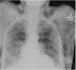
The story starts in the way you might expect from my initial preamble; a bleep for the anaesthetic team to quickly review a patient on the medical ward. The patient is an elderly lady who has had a recent chest drain inserted for drainage of her pleural effusion. She had a few other minor medical conditions including a touch of COPD but was otherwise in pretty good shape. The drain had been in and working for just over 48 hours but the day prior she had started to develop some features of surgical emphysema in her ipsilateral chest wall. More concerning for the purpose of this story is that in the past few hours she had developed a rapid spread of this up into her neck and face, with new symptoms of neck tightness, an increased sense of difficulty breathing and some voice change. The description over the phone sounded rather disturbing and so we exchanged our still warm Nescafe for our RSI grab box and headed for the lift.
On our arrival the situation wasn't quite as bad as we feared. The patient was very much controlling their own airway and, though there was a definite hoarseness to her voice, there was no stridor, no features of respiratory distress and the rest of B, C, D and E were more than satisfactory. The extent of the subcutaneous emphysema was even more marked than anticipated however. There was major swelling of the patient's neck, face, eyelids and chest wall, to the extent that eye opening was difficult.
So, what to do now? Previously I've always seen surgical emphysema as more of a cosmetic nuisance type side effect of certain procedures and something that would be left to settles spontaneously once the initial problem was corrected. But as for a definitive management when the problem was an imminent threat to life, we didn't really know what the options were. Similarly it was clear that pre-emptive measures that might be employed in other scenarios when there was the risk of imminent airway loss (e.g. smoke inhalation) was a bad idea here. If this had developed so quickly spontaneously, positive pressure ventilation was not the next best step.
Discussion
The scenario we were left with was one where we had an potential threat to airway integrity which we would likely make worse if we intervened to secure the airway. This left us with trying to improve the clinical scenario without resorting to intubation too prematurely. As such, there were a few things going through our mind at this time.
The Primary Problem
Subcutaneous (surgical) emphysema refers to the presence of air within the subcutaneous tissue planes (1,2). It can be a complication of surgical procedures (e.g. chest drains), barotrauma, or a herald of viscus/organ perforation (e.g. airway trauma, oesophageal rupture) amongst other rarities. Management rests on the identification of the cause and appropriate management of this, with ultimate resolution of the ephysema with time. Administration of supplemental oxygen has been described to help denitrogenate the subcutaneous air and therefore facilitate re-absorption. Given the relatively low incidence of the need to intervene further, any further steps are venturing into the realm of case reports/case series, which is where our path takes us.
A quick consultation with Dr Google brought up handful of similar cases with a combination of airway, breathing and circulatory failure secondary to cases of 'tension subcutaneous emphysema'. Basically, the subcutaneous air reached such a pressure that it led to either airway oedema and obstruction, chest wall rigidity and ventilatory failure, or venous return obstruction with circulatory compromise. The options available therefore involved decompressing this tension, of which a few different approaches were described.
A subcutaneous drain is one such approach, and indeed is the approach that was advised over the phone by the cadiothoracic surgeons (unfortunately at a different hospital and dealing with bleeding aortas when we were asking for advice)(3,6). This approach makes sense when you draw parallels to a pneumothorax, basically allowing an escape route for all that compressed air to escape. The techniques described may be easy enough for those with surgical experience (what’s a fascia again?), but my first thoughts were that it sound rather challenging to insert a large bore chest drain into the correct space without a high margin for misplacement (though I suspect to volume of air there is likely to be pretty large in this scenario).
An alternative 'Blow Hole' approach (4,5) more closely mirrored the management of the tension pneumothorax, and seemed to me a more practical approach for the none-surgical trained anaesthetist in a bit of bother with an impending airway disaster. After a quick bit of local to the skin the authors described making decompression incisions bilaterally at a level 3cm below to midclavicular line. The incisions were 3 cm long and down to the level of the pectoral fascia (where you'd likely get a nice hiss of escaping air). They described it as simple, quick, and in a relative safe location for the novice in a bad situation. The results they described in their case series sounded very similar to the immediate improvement that you see with needle decompression of a tension pneumothorax and avoiding the intubation that was planned.
The Backup Plan
The second major question that was on our minds related to the plan for securing an airway if everything went a bit south. The pathology present meant there was little in the way of good news here, especially when combined with an airway that was probably already a slightly challenging one anyway (though pretty hard to guess given the degree of swelling). Mouth opening was limited, Mallampati score was a 3 at best and there was nothing in the way of palpable neck landmarks. Standard direct laryngoscopy after an IV induction was clearly a bad idea here, though potentially necessary if the deterioration was sudden, and in this case the plan was to try a small size 6.0 ID tube as a first choice. An awake fibre-optic intubation or gas induction were both possible options to try and transition airway control in the safest manner, but these would still be challenging given the pathology and would have to be at least semi-elective. The final option was for an elective tracheostomy under local anaesthetic, as this was definitely not going to be a plausible rescue technique. Neither of the options had particularly developed as a favourite in our minds, but fortunately that particular bullet was dodged.
So how did our story end? Well as I alluded to earlier, the patient wasn't immediately peri-arrest and, despite some worrying features, there was no suggestion of imminent airway loss. The balance of risk vs benefit of early intubation and PPV was still on the risky side of the spectrum and favouring active treatment of the emphysema in the first instance. On the advice of the cardiothoracic team the medical team had successfully inserted a drain into a roughly subcutaneous location and some active massaging of the tissue in a head to feet direction produced some air escape, mostly out of the chest drain. Indeed the patient felt enough of an improvement in symptoms for us to feel comfortable getting a CT scan for some further information (demonstrating a persistent pneumothorax and subcutaneous air eveywhere). Insertion of a second chest drain was carried out and the patient was transferred to the high dependency unit for close observation. Fortunately there was progressive resolution of the emphysema over the next few hours, correlating well with decreasing anaesthetic staff stress levels.
Final Thoughts
Overall an interesting challenge that asked a lot of different questions of us. I think a reflection such as this should finish with final thoughts about how we would manage the case if we had it a second time. Here are my thoughts:
1. Watchful waiting was almost certainly a better option than pre-emptive intubation in this case give the patient’s stability and the risk of worsening things with PPV. Unless absolutely no choice, I’d try avoid the tube next time. This is quite different from other cases of an at risk airway (e.g. smoke inhalation).
2. If my hand was forced the described subclavicular decompression technique seems a relatively safe last throw of the dice to try and rescue an imminent airway/breathing/circulation failure if help can’t arrive in time. Using the relevant cardiothoracic or even ENT surgical help would be the ideal scenario if time permitted, but having a rescue technique in the back of my head feels comforting.
3. I think either an awake tracheostomy or fibre-optic intubation would be the most comfortable out of all the options available, though both of these would require a degree of time and therefore would probably come after the attempt at decompressing the neck.
Thanks for reading and I hope it was of interest. As always I’d love to hear your thoughts, especially if you have had the challenge of similar scenarios in the past as I think there is a wide scope for different approaches here which are worth discussing. I also recommend checking out the cases below if you haven’t already for some more detail on the topic.
BW
Tom Heaton
References
- Medscape - Air Leaks, Pneumothorax and Chest Drains
- Radiopaedia - Subcutaneous Emphysema
- Case Report; Subcutaneous Drain. Anaesthesia. 1995
- Case Report: Life Threatening Subcutaneous Emphysema. Anaesthesia. 2008
- 'Blow Holes' Management. Chest. 1992
- Case Report: Subcutaneous Drain. Respirology Case Reports. 2013
- Upper Airway Obstruction Case. BJA. 2004
Image courtesy of radiopaedia.org
 RSS Feed
